I.trimester
I.trimester
First trimester screening is performed when the fetus is between 11 weeks + 3 days and 13 weeks + 6 days old, with a crown-rump length (CRL) of 45-84 mm. At this stage of pregnancy, it is possible to diagnose certain congenital organ defects and assess so-called ultrasound markers for fetal diseases. An important feature is the swelling of the skin in the neck area (nuchal translucency – NT). Significant enlargement of NT may indicate a risk of congenital defects. It is also important to assess other signs such as the presence of the nasal bone (NB), tricuspid regurgitation, and ductus venosus. This examination also includes a maternal blood test to determine the levels of placental hormones and proteins, PAPP-A and free βhCG. Based on these examinations, the combined risk of chromosomal abnormalities for the fetus, especially Down, Edwards, and Patau syndromes, is determined.
YOU CAN HAVE THE SCREENING RESULT AT OUR DEPARTMENT ON THE SAME DAY.
| Combined first trimester screening including preeclampsia – genetic risk assessment – singleton pregnancy | 2 000 Kč |
| Combined first trimester screening including preeclampsia – genetic risk assessment – twins | 3 000 Kč |
Preeclampsia screening
Preeclampsia is a condition that occurs only during pregnancy and is characterized by a triad of symptoms – increased blood pressure, significant protein loss in the urine, and generalized swelling. This condition manifests after the 20th week of pregnancy and in the most severe cases can lead to the induction of preterm birth or termination of pregnancy by cesarean section, which can be associated with serious complications for the newborn. Unfortunately, it can also pose a threat to the health and life of the mother. The test to determine the risk of preeclampsia is performed simultaneously with the combined first trimester screening and has a detection rate of up to 90%. Performing this test is recommended for all pregnant women.
The test has three parts:
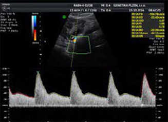
- Ultrasound examination of blood flow in the uterine arteries by a certified sonographer (certificate from FETAL MEDICINE FOUNDATION London)
- Repeated blood pressure measurement on both upper limbs according to a precisely defined procedure
- Blood test of the pregnant woman to determine the level of placental growth factor (PLGF).
In case of increased risk of preeclampsia, preventive treatment is recommended to the pregnant woman, which significantly reduces both the likelihood of occurrence and the severity of the potential condition.
II. trimester
II. trimester
An ultrasound examination performed between week 19 and 22 of a woman's pregnancy. It is the most important ultrasound examination to exclude congenital defects of foetal organs. Apart from evaluating the foetus as a whole and each organ, we also determine foetal weight and therefore the correct growth of the foetus. Last but not least, we evaluate the position and appearance of the placenta and the amount of amniotic fluid.
III. trimester
III. trimester
Examination in week 30-32 of a woman's pregnancy. In this period of time the key examination includes the size and position of the foetus. The organs are again evaluated, including the status and position of the placenta and the amount of amniotic fluid. In some cases we measure blood flow rate in the umbilical cord and other foetal vessels.