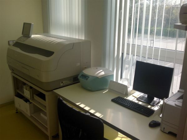
Our laboratory was founded in 2003. Since that time we have continued to expand both in terms of personnel and equipment as well as the spectrum of performed methods.
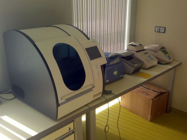
Due to the quality of the instruments in our laboratory we are able to perform molecular genetic examinations in accordance with the latest diagnostic trends. We carry out detection of known mutations using PCR, or Real Time PCR and detection of mutations using direct sequencing to diagnose monogenic hereditary diseases. We also perform quick diagnostics of the most common aneuploidies from native amniotic fluid using multiplex QF PCR. We ensure transfer of the samples for DNA examination of various hereditary diseases to various departments in the Czech Republic, and if needed abroad.
We can offer determination of paternity (or maternity, parity) including expert opinions. The expert opinions are done at the request of the court, authorities involved in criminal proceedings and private persons.

Our laboratory is accredited according to ČSN EN ISO 15189.
We successfully take part in annual external quality audits.
Dokumenty
List of performed methods
QF PCR (Quantitative Fluorescent Polymerase Chain Reaction) testing serves as a rapid diagnostic tool for the most common aneuploidies and uniparental disomy of autosomal chromosomes (13, 18, 21, 15, 16, 22) and sex chromosomes (X, Y). This involves the analysis of specific STR loci using PCR and subsequent fragment analysis through capillary electrophoresis.
Detection of hereditary diseases primarily includes Down, Edward, Patau, Klinefelter, Turner, Jacob (supermale, XYY), triple X (superfemale, XXX) syndromes, and polyploidies.
Aneuploidy testing can be performed on amniotic fluid, chorionic villi, fetal blood, and miscarried fetal tissue. It is followed by cytogenetic analysis of all chromosomes.
Indications for testingpositive atypical or borderline first and second trimester screening, overall genetic risk for the fetus above 10%, abnormal ultrasound findings, combination of multiple risk factors.
Cystic fibrosis (mucoviscidosis) is a multisystem autosomal recessive genetic disorder that, in its classic form, manifests as respiratory tract and lung disease, pancreatic exocrine insufficiency, high sweat electrolyte levels, and male infertility. It is caused by mutations in the CFTR gene. The incidence in the Czech population is 1:4500, with a carrier frequency of 1:34 (Votava F. et al., 2014).
Our laboratory tests for 68 mutations in the CFTR gene and determines polymorphism in intron 8 (Tn variants). The mutations tested represent 96% of the causal mutations in the CFTR gene in the Czech population. Excluding these mutations reduces the probability of carrying a mutation in the CFTR gene to 1:847 (0.118%). The test can detect both asymptomatic carriers (heterozygotes) and affected individuals (homozygotes). Here you can find the List of Reference Sequences.
Indications for testingpatients with symptoms of cystic fibrosis, relatives of a patient with cystic fibrosis and detected CFTR mutations, partner of a mutation carrier before planned pregnancy or during pregnancy, adult males with infertility issues, prenatal diagnosis in case of partners heterozygous for a CFTR gene mutation, preconception screening, gamete donors.
Analysis of the AZF region (azoospermic factors) on the long arm of the Y chromosome (Yq) is divided into AZFa, AZFb, and AZFc. The genes located in this region are involved in spermatogenesis and are essential for male reproduction. Depending on which azoospermic factor is missing, men may experience oligozoospermia (reduced sperm count in ejaculate) to azoospermia (absence of sperm in ejaculate). Testing of STS loci on Yq detects approximately 90% of deletions in the AZF regions.
Indications for testing: male fertility disorder - poor spermiogram parameters
The test is conducted to detect the c.657-661del mutation, a deletion of five nucleotides, in the NBN gene. This mutation, in a homozygous state, is responsible for Nijmegen breakage syndrome (NBS), also known as Seemanová syndrome. NBS is a rare autosomal recessive DNA repair disorder. It is a chromosomal instability syndrome characterized by microcephaly, growth retardation, immunodeficiency, and a predisposition to malignancies. An increased incidence of malignancies also affects heterozygous carriers. Patients with NBS are hypersensitive to ionizing radiation. This mutation is more common in the Slavic population, with a carrier frequency of 1:100-150.
Indications for testingsuitable for patients with clinical manifestations of microcephaly, chromosomal instability, growth retardation, increased frequency of malignancies in family history (especially lymphomas and leukemias), reduced levels of IgG and IgA in serum.
Hemochromatosis is a disorder of iron storage that results in its accumulation in parenchymal cells. As the disease progresses, it leads to extensive tissue damage, diabetes mellitus, liver cirrhosis, hepatocellular carcinoma, heart failure, arthritis, and pigmentation. The first symptoms often appear between the ages of 40 and 50. It is an autosomal recessive disorder.
We detect the most common mutations c.845G>A (C282Y) and c.187C>G (H63D) in the human HFE gene using allele-specific PCR. The test can detect both asymptomatic carriers (heterozygotes) and affected individuals (homozygotes).
Indications for testing: liver fibrosis or cirrhosis, cardiomyopathy, pancreopathy, diabetes, arthropathy, skin pigmentation.
Thrombophilia is a tendency toward increased blood clotting, where there is an elevated risk of blood clot formation (thrombi), which can lead to partial or complete blockage of blood vessels, most commonly in the lower limbs. A part of the clot can dislodge, travel through the venous system, and after passing through the heart, block one of the pulmonary vessels, resulting in a pulmonary embolism. We examine two mutations in the F5 and F2 genes. These genes encode the proteins factor V and factor II, which play a significant role in the blood clotting process. The mutation in the F5 gene is called Leiden (FV Leiden – G1691A). In the Czech population, 5-10% are carriers of this mutation in a heterozygous state (one mutated allele), which increases the risk of thromboembolic disease approximately 7-fold. In the case of homozygotes (both alleles mutated), the risk increases about 20-fold. The second mutation is the Prothrombin mutation in the F2 gene. Heterozygotes in the population are approximately 2-3% and increase the risk of thrombosis about 3-fold. In homozygotes, it is up to 18-fold.
Indications for testing:
- venous thrombosis (embolism) in an individual under 50 years of age
- thrombosis occurring in an unusual body part
- thromboembolic disease in a patient with a family history of thromboembolism
- thromboembolic disease in a patient with a family history of thromboembolism
- patient prior to scheduled surgery with a family history of thromboembolism
- a patient with recurrent miscarriages in the second or third trimester without a clear cause
- indication by a clinical haematologist
Congenital hearing disorders occur in approximately 1 in 1000 newborns, and about half of these are genetically caused. Most often, it is an autosomal recessive nonsyndromic (isolated) hearing loss. We examine prevalent mutations – c.35delG (p.G12fs) and c.71G>A (W24X) in the connexin 26 gene (GJB2), which are the most common cause of nonsyndromic hearing loss. The test can detect both affected individuals (homozygotes) and hearing carriers (heterozygotes).
Indications for testing:
- prelingual hearing loss
- prelingual hearing loss in the family
- congenital hearing loss prior to cochlear implantation
- the presence of the mutation in consanguineous partners, or in newborns of incestuous relationships
- hearing relatives in families where the mutation has already been detected
Celiac disease is an autoimmune disorder caused by intolerance to gluten, which is a protein component of wheat, rye, and barley. The disease causes inflammation of the small intestine's mucosa and destruction of villi and microvilli. The most common symptoms are abdominal pain, bloating, diarrhea, occasionally constipation or vomiting, loss of appetite, and weight loss. In young children, a distended abdomen and growth disorders can often be observed.
During testing, we detect HLA alleles predisposed to celiac disease. The alleles being tested are found in more than 99% of patients with celiac disease and in 20% of healthy controls. The presence of predisposed alleles increases the risk of developing celiac disease 50 times compared to the general population.
Indications for testing:
- failure to thrive
- indigestion
- occurrence of celiac disease in the family
- anemia of unknown origin
Bechterew's disease (ankylosing spondylitis) is a chronic inflammatory disease primarily affecting the spinal vertebrae. The exact cause is unclear, but it has been found that more than 95% (in the Caucasian population) of patients with ankylosing spondylitis have the HLA-B27 antigen. The occurrence of the HLA-B27 allele in Caucasians is about 8%. Individuals with the HLA-B27 antigen have a probability of developing the disease up to 300 times higher compared to those who do not possess this antigen. However, the presence of the HLA-B27 antigen does not necessarily mean that the disease will develop. It only indicates an increased risk.
In the test, we detect the presence of the HLA-B27 allele. It is not suitable to use this test for screening the asymptomatic population for the detection of ankylosing spondylitis, but the test indicates an increased likelihood of occurrence in a symptomatic patient. The test is also suitable for ruling out the disease in a clinically unclear patient.
Indications for testing:
Back pain, chronic pain, or stiffness in the lower spine, reduced spine flexibility, chest pain during deep breathing, chronic fatigue, inflammation of the iris and ciliary body (iridocyclitis), less common symptoms include joint inflammation, pulmonary fibrosis, and nail separation from the nail bed.
Fragile X Syndrome (FraX) is an X-linked disorder characterized by mental retardation with possible dysmorphic features (elongated face with prominent chin and large protruding ears). The syndrome is caused by a dynamic mutation – expansion of CGG trinucleotides in the 5' untranslated region of the FMR1 gene. The number of repeats varies from the normal number of repetitions, through an intermediate mutation, premutation, to a full mutation.
Carriers of the premutation are not affected by mental retardation, but the premutation causes, with low penetrance, presenile tremor and premature ovarian failure in women (approximately 20% of female premutation carriers). Premutations are unstable during meiosis or early embryogenesis, and if transmitted by a woman, there is a risk of CGG-repeat expansion to a full mutation. In contrast, premutations transmitted by men rarely expand to a full mutation. Mental retardation caused by the full mutation primarily affects men, but the disease can also manifest in women. Approximately half of the female carriers of the full mutation have mild to moderate mental retardation.
During the PCR test, we determine the number of CGG repeats in the FMR1 gene.
Indications for testing:
- psychomotor retardation, autism in personal or family history
- premature ovarian failure
- presenile tremor in men
Gilbert's syndrome is a lifelong autosomal recessive hereditary disorder of bilirubin metabolism in the blood. It is caused by a dysfunction of UDP-glucuronyltransferase 1A1, reduced to 20-30% in affected homozygotes. It most commonly manifests as mild hyperbilirubinemia without signs of liver disease or hyperhemolysis. The condition is considered to be of low severity. Most often (over 90% of cases), the cause is an increase in the number of TA repeats in the promoter TATA box of the UGT1A1 gene from 6 to 7.
During testing, we detect the number of TA repeats in TATA, and in the case of finding a homozygous result of 7TA/7TA, we confirm Gilbert's syndrome.
Indications for testing:
- isolated mild hyperbilirubinemia (suspected GS)
- relatives with confirmed GS
- before taking irinotecan (GS represents a pharmacogenetic risk factor for irinotecan toxicity)
As in all mammals, the ability to digest lactose in humans is greatest during infancy and decreases with age. An adult produces only one-tenth of the lactase compared to an infant, and most people by the age of 60 cannot digest lactose at all. It is estimated that 75-80% of the world's population is lactose intolerant in adulthood. The most common symptoms are lactose digestion disorders, flatulence, abdominal distension, and diarrhea. Lactose intolerance varies among ethnic groups, with the prevalence around 30% in the European population. We examine the -13910C>T variant in the promoter region of the LCT gene.
The examination is suitable for both, the identification of asymptomatic carriers (heterozygotes) and the molecular-genetic diagnosis of affected individuals (homozygotes).
Indications for testing:
- gastrointestinal upset after eating foods containing lactose
- differentiation of the primary type of lactase deficiency from the secondary type
- pathological lactose tolerance test or positive H2 breath test
- osteoporosis
Non-invasive prenatal testing (NIPT) is an examination of the most common fetal aneuploidies from maternal blood. It uses free DNA (a mixture of free maternal and fetal DNA) isolated from plasma. We use the Panorama test (by Natera), which is based on targeted SNP sequencing, allowing it to distinguish between free fetal DNA and free maternal DNA. The Panorama test shows high sensitivity for trisomies of chromosome 21 (99%), 13 (99%), 18 (96.4%), and sex chromosomes (100%), as well as for monosomy of chromosome X (92.9%). It can also detect vanishing twin syndrome, triploidy, and test for aneuploidies of chromosomes 13, 18, 21 in twin pregnancies and pregnancies from a donated oocyte.
Indications for testing:
- increased genetic risk of chromosomal aberration for the fetus (chromosomes 21,18, 13, X, Y)
- normal ultrasound findings while positive/borderline/atypical prenatal screening
- increased risk of complications when performing invasive diagnostics (CVS/AMC) because of higher genetic risk
- older age of pregnant woman (over 38 years)
- pregnancy via IVF, dysfertility, or sterility treatment
Only a small proportion of tumors (5-10%) are inherited, i.e. they are hereditary tumor syndromes.
Hereditary cancers arise as a result of inherited mutations (disorders of genetic information in the DNA) of tumor predisposing genes, of which several hundred are known. The causal mutation is passed on from generation to generation in the affected family. The offspring can inherit it from the carrier in half of the cases. Its presence significantly increases the likelihood of developing cancer.
Early analysis of hereditary cancer predisposition is extremely important for high-risk individuals and allows mutation carriers to be included in preventive monitoring programs and consideration of possible prophylactic surgeries. The most common hereditary forms of cancer are breast, ovarian, and colorectal cancers.
Hereditary breast and/or ovarian cancer syndrome is an autosomal dominant syndrome with incomplete penetrance. The causes are causal (pathogenic) mutations primarily in the BRCA1 or BRCA2 tumor suppressor genes. Besides BRCA1,2 genes, genes such as TP53, STK11, CDH1, PTEN, PALB2 are considered high-risk concerning breast cancer, while genes ATM, CHEK2, NBN are considered moderate-risk. For ovarian cancers, high-risk genes for Lynch syndrome (MLH1, MSH2, MSH6, PMS2) as well as BRIP1, RAD51C, and RAD51D genes are considered.
Colorectal cancers can be divided into non-polyposis – Lynch syndrome (genes MLH1, MSH2, MSH6, PMS2, EPCAM, MUTYH) and adenomatous polyposis – FAP (APC gene).
In addition to breast, ovarian, and intestinal tumors, heredity can also apply to tumors of the stomach, skin, pancreas, kidneys, and others.
Mutation analysis/screening of these genes allows confirmation of the genetic background of cancer development in affected individuals and subsequent predictive testing of their biological relatives. Asymptomatic mutation carriers are included in a dispensary program aimed at early detection of possible cancer. For testing, we use the Czecanca panel (Czech cancer panel for clinical application), developed by the team of doc. MUDr. Kleibl from the Institute of Biochemistry and Experimental Oncology, First Faculty of Medicine, Charles University in Prague. The panel contains 226 target genes for the investigation of hereditary malignancies using Next Generation Sequencing (NGS) – here you will find a List of Reference Sequences. It allows analysis of all known predisposition genes (approximately 69 genes) whose mutations are the cause of hereditary cancers (e.g., genes ATM, APC, BARD1, BRCA1, BRCA2, BRIP1, CDH1, CHEK2, EPCAM, MLH1, MSH2, MSH6, MUTYH, NBN, PALB2, PMS2, PTEN, RAD50, RAD51C, RAD51D, STK11, TP53, CDKN2A, CDK4, and many others). Other genes included in the panel represent candidate genes for preclinical studies within the Czech population.
Indications for testing:
- cancer at an unusually young age
- people with multiple types of cancer
- occurrence of bilateral tumors in paired organs
- recurrence of the same type of cancer in a family history (e.g. breast or colorectal cancer)
- combination of certain types of tumors in close relatives (breast and ovarian cancer, colorectal cancer and uterine cancer), especially in cases where the disease manifested at a younger age
Spinal muscular atrophy (SMA) is the second most common cause of death in children due to an autosomal recessive disease (incidence of 1 in 10,000 live births). The frequency of heterozygotes, i.e., healthy carriers of the disease, in the population is 1:47.
SMA is an autosomal recessive neuromuscular disease characterized by the degeneration of spinal horns. The cause is a mutation in the SMN1 gene. In more than 95% of patients, it involves a homozygous biallelic deletion of SMN1 detected using exon 7. Besides the SMN1 gene, there is a highly homologous pseudogene SMN2 on the same chromosome (Chromosome 5). Most of its transcripts, due to the 840C>T substitution, lack exon 7. The protein produced by this gene is insufficient to prevent the disease but is enough to modify the phenotype. Determining the number of SMN2 gene copies is significant for SMA patients because the more copies a patient has, the milder the symptoms of the disease.
We perform the examination using the MLPA method to determine large deletions and duplications, which allows us to detect the number of copies of exon 7 and 8 in the SMN1 gene and the SMN2 pseudogene.
Indications for testing:
- gamete donors
- preconception examination
- individuals with neuromuscular disease
- families with SMA and relatives of already diagnosed SMA carriers